
Mushroom Cap Shapes: Convex, Bell, Flat, and Funnel Clues
Learn which mushroom cap shapes a photo can show, which clues are reliable, and the safe next steps to compare convex, bell, flat, and funnel caps.
Quick answer for mushroom cap shapes
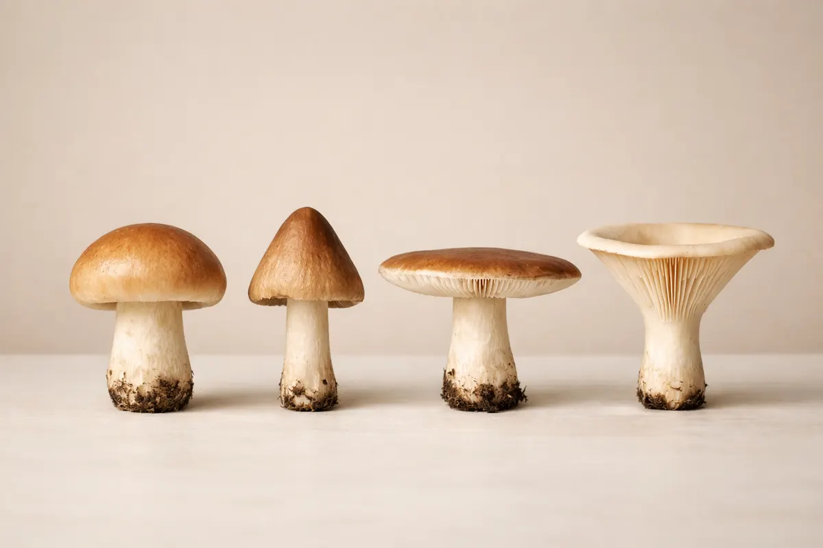
Mushroom cap shapes like convex, bell, flat, and funnel are among the clearest features a photo can record — but a single image rarely proves species identity on its own. Cap shape gives a strong initial filter: a clearly funnel-shaped cap excludes many gilled mushrooms, while a persistent bell shape narrows possibilities among young agarics and some small boletes. Treat cap shape as one axis in a checklist, not as a final label.
When you inspect a phone photo for cap shape, the most reliable signals are overall silhouette and margin behavior. Is the cap bowl-like with the center depressed, or rounded and dome-like? Does the edge roll under, remain straight, or lift and flange outward? These observations survive modest blur and imperfect lighting better than color or subtle texture.
Use cap shape to short-list likely groups, then combine that short-list with other clues — gill attachment, pore surface, stem attachment, habitat, and spore print when possible. If you want a fast next step, take additional images that show the underside, stem base, and a top view with scale; these will quickly confirm whether a convex cap is simply a young flat cap or a genuinely bell-shaped species.
- Cap shape is a strong first filter but not proof of species.
- Silhouette and margin behavior are the most photo-stable clues.
- Follow-up photos (underside, stem base, scale) turn a shape clue into a practical shortlist.
Strongest visual clues for identifying cap shapes
Start with the cap outline: a convex cap has a rounded dome, a bell cap appears conical with a flared skirt at the margin, a flat cap is broadly planar often with a slight central bump, and a funnel cap has a central depression that deepens to form a throat. In phone photos, that overall outline — especially when you have a top or three-quarter view — resists small lighting changes and is the most dependable descriptor of cap shape.
Next, check the margin behavior. The cap edge (margin) tells a lot: incurved margins that later uncurl indicate a young convex cap maturing toward flat; margins that are thin and pendulous point toward bell-shaped species; margins that curve upward and create a visible flange often signal old or flattened caps. Margins are visible even in partially obscured or dappled-light photos and help separate lookalikes that share similar dorsal textures.
Center structure and surface slope are also durable. A distinct central umbo (a raised bump) or a depressed center tends to persist across multiple photos and angles. For example, many russulas and some agarics present a convex cap with a clear central bump, whereas certain chanterelles and Craterellus species show a shallow, wide funnel. Pay attention to how the cap slopes away from the center — steep slope, gentle dome, or a gradually depressed throat.
Texture and persistence under touch are weaker than outline but can reinforce shape clues. Radial striations, scalable patches, and the presence of a pronounced margin (skirt) line are photosensitive details that often survive low resolution. Combine texture cues with outline and margin to raise confidence: a bell-shaped cap with a smooth, striated margin suggests a different set of genera than a bell cap with scaly fragments.
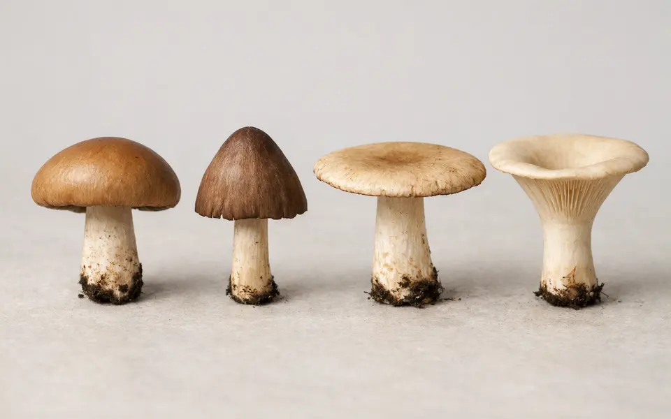
- Outline/silhouette: primary durable clue for convex, bell, flat, funnel.
- Margin behavior (incurved, straight, uplifted) helps distinguish age vs. species effects.
- Center features (umbo, depression) are visible across angles and support shape calls.
- Surface texture and striations add supporting evidence when present.
Weak signals to treat cautiously when reading cap shapes
Color and subtle shading are unreliable for cap shape assessment because they change with light, moisture, and age. For example, a damp convex cap darkens and can appear flatter in photos; a sun-bleached bell cap may lose its visible skirt and be mistaken for a different shape. Rely on color only to hint at moisture or age, never as the primary cap-shape indicator.
One-angle photos and strong glare can mislead. A single top-down image hides margin curvature and underside features; a side-only image might exaggerate dome depth or hide a shallow funnel. Glare and specular highlights can remove visual cues about slope and texture, so flag photos with strong reflections as low-confidence for cap shape.
Small adults and juvenile stages are common pitfalls. Many mushrooms start convex and flatten with age; several bell-shaped species begin as hemispherical buttons. If the specimen looks small relative to nearby twigs or leaves, note the possibility it’s an immature stage. Size without scale is itself a weak signal: always include a coin or ruler when possible for decisive comparison.
Environmental distortion matters. Wind-bent, waterlogged, or insect-chewed caps can lose their normal silhouette. Likewise, clusters or overlapping caps create composite outlines that obscure individual shapes. Where multiple fruiting bodies grow together, isolate a single mature specimen before assigning a shape.
- Avoid treating color as proof of shape — it’s influenced by moisture and light.
- Single-view photos and glare reduce confidence in margin and center features.
- Juvenile size and growth stage often mimic other cap shapes — confirm age or include scale.
- Clustered or damaged caps can hide the true silhouette; isolate one specimen where possible.
How to compare likely matches using multiple cap-shape clues
Comparison is a process of elimination: use cap shape to create a shortlist, then test each candidate against other independent clues. Start by writing two or three shape descriptors for your specimen (for example: convex with central umbo; margin slightly flared; underside appears gilled). Each descriptor becomes a filter when you scan field guides or image searches: discard species that cannot display that combination even at different life stages.
Match margin behavior next. If your cap shows an incurved margin in the photo, prefer species known to start with incurved edges. If the margin is strongly uplifted, consider older caps or species with naturally uplifted margins like many chanterelles and Craterellus relatives. Use this filter to separate species that share a rounded silhouette but differ in edge morphology.
Bring in underside structure early. While cap shape narrows groups, whether the underside has gills, pores, ridges, or folds is often the deciding characteristic. A funnel cap with decurrent ridges points you toward chanterelles (Cantharellus/Craterellus) rather than gilled agarics, even if the dorsal shape looks ambiguous in a crowded photo.
Use habitat and substrate as tie-breakers. Some cap shapes are common across habitats, but their occurrence on wood, leaf litter, or grass can exclude many lookalikes. For example, a flat, thin-capped agaric on lignin-rich wood is probably a saprotrophic wood-decomposer rather than a mycorrhizal bolete with a similar top view. Combine cap shape, margin, underside, and substrate in sequence to rank likely matches from most to least plausible.
Keep a short, explicit uncertainty note with each comparison. Jot down why a candidate was kept or rejected (e.g., “kept: convex with umbo and free gills; rejected: funnel because underside shows pores in observed photo”). That audit trail makes it easier to revisit the decision after collecting an extra photo or a spore print.
- Step 1: Describe the cap using 2–3 durable traits (outline, margin, center).
- Step 2: Filter candidates by margin behavior and likely life stage.
- Step 3: Check the underside (gills, pores, ridges) to rule groups in or out.
- Step 4: Use substrate and habitat as decisive context cues.
- Step 5: Record brief reasons for keeping or rejecting each candidate.
Practical app workflow after checking cap-shape clues
Once you’ve noted cap shape and other immediate clues, use an identification app as a structured next step rather than a final authority. On your phone, open Mushroom Identifier: Fungio (or your preferred app) and feed it multiple photos: a top view showing the cap outline, a clear side view showing margin and slope, a close-up of the underside, and a picture of the stem base and substrate. Multiple angles let the app combine visual cues in the same way you would during manual comparison.
Enter short field notes into the app’s description field: cap shape (convex/bell/flat/funnel), margin behavior, substrate type (wood/leaf litter/grass), and whether the specimen is in a cluster. These text clues help the app prioritize visual matches and create a more useful shortlist for your review. Keep your uncertainty visible: mark whether the specimen looks juvenile or weathered so the algorithm treats those matches differently.
Treat app results as hypotheses to test, not conclusions. Use the app’s candidate list to pull up high-quality reference images and look specifically for the features you recorded: does the candidate show the same margin behavior and underside structure? If app suggestions disagree, return to the comparison workflow: rule out candidates that fail any one durable clue, and collect a spore print or a scale photo if necessary.
If you want to document your process for later review or expert consultation, save the photos and your short notes. That record lets a more experienced identifier or a mycological forum check your reasoning quickly and suggest precise follow-ups — for example, a spore print color test or a microscopic examination — rather than asking for the same basic views again.
- Upload or scan multiple photos: top, side, underside, stem base (use phone camera).
- Add brief field notes: cap shape, margin, substrate, and age/condition.
- Use the app’s candidate list to confirm durable clues, not to finalize ID.
- Save images and notes so experts can review your reasoning and suggest next checks.
Related guides
After you check cap-shape clues, try Mushroom Identifier: Fungio
Use Mushroom Identifier: Fungio as a structured next step—scan multiple photos (top, side, underside, stem base) and add your short notes about cap shape, margin behavior, and substrate. Treat the app’s suggestions as hypotheses to test: compare candidates against the durable clues you recorded and save your images and notes for follow-up or expert review. Visit https://mushroomidentification.app/ to open the app and continue the checklist.
Take the next step →Frequently asked questions
Can cap shape alone identify a mushroom species?
No. Cap shape is a powerful filtering clue but not sufficient for species-level identification. Many unrelated species share convex, bell, flat, or funnel outlines at some life stage. Always combine cap shape with underside structure (gills, pores, ridges), stem features, habitat, and, when feasible, spore print color. Treat a shape-based ID as a working hypothesis that requires at least one independent confirming clue.
How can I tell if a mushroom is bell-shaped or just a young convex cap?
Compare margin behavior and maturity signs: bell-shaped species often retain a distinctly pendulous or flared margin and sometimes a thin, skirt-like edge even when mature. Young convex caps typically have margins that are incurved and later uncurl as the cap flattens. Look for multiple specimens at different stages: if all show a sustained conical or skirted margin, bell shape is more likely. When in doubt, photograph margin detail and underside to confirm.
What photos should I take to confirm cap shape and avoid mistakes?
Take a top or three-quarter view to capture the overall silhouette, a side view that includes the cap margin and stem, a close-up of the underside to show gills or pores, and a picture of the stem base and substrate for ecological context. Include a scale object like a coin or ruler when safe. If the cap is in a cluster, isolate a single mature specimen for these shots so overlapping forms don’t confuse the outline.
Which lookalikes most commonly confuse funnel, bell, and flat caps?
Common confusion pairs include young flat-capped agarics mistaken for convex domes, collapsed or weathered caps that appear funnel-like but belong to gilled species, and clustered chanterelles versus small funnel-form Craterellus species. Damage, age, and moisture collapse disguise true silhouettes. Use underside structure (ridges vs gills vs pores) and substrate to separate these lookalikes reliably.