
Mushroom Visual Checklist: Common Clues, Lookalikes, and Next Steps
A practical mushroom visual clues checklist: what to look for in photos, common lookalikes, and safe next steps to verify uncertain matches before you act.
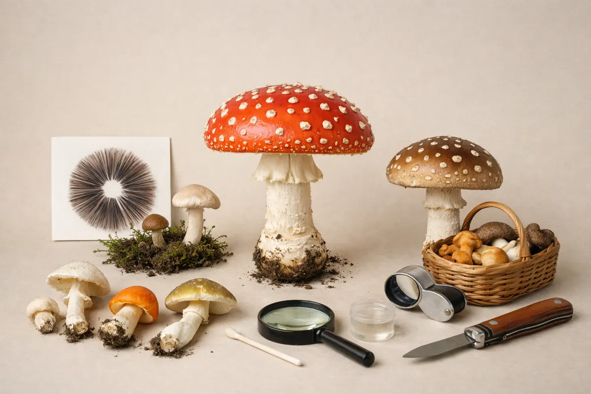
Quick answer for mushroom visual clues
Mushroom visual clues are the observable features—cap shape and color, gill or pore structure, stem details, surface texture, bruising, and the surrounding habitat—that let you narrow which species are possible from a photo.
A single image can often rule out many families but rarely proves edibility or toxicity. Treat an on-phone visual checklist as a first-pass filter: collect multiple angles, note habitat and substrate, and then use a step-by-step verification plan before drawing conclusions.
- Look at cap shape, margin, surface texture, and size.
- Check gills, pores, or spines from underneath and note attachment to the stem.
- Document stem base and any volva, ring, or rooting structure; record habitat (wood, leaf litter, lawn).
What it means
When people search for mushroom visual clues they want reliable, photo-based hints that reduce thousands of fungal species to a manageable shortlist. Visual clues are the field marks used by mycologists the moment they see a specimen: cap form (convex, bell, flat, funnel), underside structure (gills, pores, teeth), and stem features (ring, volva, texture).
Visual clues include secondary signals as well: where the mushroom grows (on wood, turf, dung), whether fruitbodies are solitary or clustered, and how they age or bruise. Taken together these data points create a pattern that narrows possibilities from family to genus and sometimes to species.
A photo-based checklist is useful because many lookalike groups separate cleanly by a few visible traits. For example, chanterelles (Cantharellus spp. ) have blunt, forked ridges and a fruity smell, while true jack-o'-lanterns (Omphalotus spp. ) have true gills and grow on wood. Those contrasts are visible even in good phone photos.
However, some important diagnostic features require touch, smell, spore color (spore print), or microscopy; visual clues are rarely the whole story. Use photos to prioritize next steps—more images, spore print, habitat notes, and expert consultation—not to confirm safety.
Key clues
Below are high-value visual clues that are easy to check with a camera and often decisive for narrowing lookalikes. When you photograph a specimen, aim to capture these features clearly and include a scale (coin or ruler) in at least one frame.
Each bullet includes why that clue matters and what to look for in photos.
- Cap shape and margin — Note whether the cap is convex, bell-shaped, umbonate (with a central bump), depressed, or funnel-shaped. Margin details (inrolled, wavy, striate) are often species-specific.
- Cap surface and texture — Smooth, viscid (slimy when wet), scaly, fibrillose (silky strands), or cracking surfaces point to different genera. Color changes with age or when bruised are important to document.
- Underside structure — Gills vs. pores vs. teeth (spines) is among the single most useful clues. Also photograph gill spacing (crowded vs. distant), color, and whether gills attach to, run down, or stop before the stem.
- Stem details — Measure thickness, look for a ring (annulus), volva (cup at the base), rooting base, or surface ornamentation like fibrils or granules. A swollen or bulbous base often separates toxic Amanitas from lookalikes.
- Attachment and context — Are fruitbodies solitary, in clusters, or in rings? Growing on wood, leaf litter, turf, or dung drastically changes likely species.
- Bruising and color change — Rub the cap or stem (gently) and watch for immediate color changes or delayed bruising over minutes. Some boletes bruise blue; others will not. Photograph before and after bruising.
- Spore print color — While not purely visual in-field, a spore print (lay cap gills-down on white and dark paper for several hours) produces a crucial color clue: white, brown, black, pink, or olive. Include the method and timing in notes.
- Odor and exudates — Smell notes (mild, fruity, unpleasant) and any droplets or latex on cut surfaces can be diagnostic for genera like Lactarius or Hygrophoropsis.
Step-by-step workflow
Turn visual clues into a clear verification workflow you can follow every time. This reduces guesswork and creates a record you can share with a local expert or an app without overclaiming certainty.
Keep a short checklist on your phone so you don’t miss frames: cap top, cap edge, underside close-up, stem mid-section, stem base (dig gently around base), habitat wide shot, and a scale reference.
- Step 1 — Photograph systematically: 1) cap top centered, 2) cap margin close enough to see texture, 3) underside (gills/pores/teeth) in good light, 4) full mushroom showing scale and habitat, 5) stem base after carefully exposing it.
- Step 2 — Record context: note substrate, tree species nearby, number of fruitbodies, date/time, weather, and any smell or exudate observations.
- Step 3 — Compare visible clues: check the cap shape, underside type, stem features, and habitat against a trusted field guide or illustrated keys. Narrow to a family or a short list of genera.
- Step 4 — Run simple verification checks: spore print color, bruising tests, and odor notes. These often eliminate lookalikes more reliably than color alone.
- Step 5 — Treat app or photo-ID suggestions as research notes: use them to refine your shortlist but do not assume correctness. If a result could impact safety (pets, kids, ingestion), escalate to a local mycological society, clinic, or poison control with your photos and notes.
Examples
Example 1 — White cap in a suburban lawn: A small, smooth white cap with crowded, free gills and a faint ring risks being mistaken for several groups. Key next checks: expose the stem base for a volva (Amanita warning sign), take a spore print, and note whether it grows solitary or in fairy rings. For a deep dive on white lawn species and risks, see White Mushroom in Yard: Photo Clues, Risks, and Safe Next Checks (https://mushroomidentification. app/blog/white-mushroom-in-yard).
Example 2 — Clustered orange mushrooms on rotting oak: Bright orange funnel shapes with decurrent ridges suggest chanterelle-like fungi, but clustered growth on wood points to a jack-o'-lantern lookalike. Photograph the underside carefully: true gills rule out chanterelles. Next steps include detailed underside photos and checking for growth on buried wood.
Example 3 — Large brown bolete under pines: A stout brown cap with a spongy pore surface under conifers indicates a bolete. Look for blue bruising on the pore surface and stem patterning; note whether the pores stain a different color when pressed. Boletes have many edible and inedible species; spore print and chemical tests help, but consult an expert for anything beyond confident, well-known edibles.
Limitations
High-confidence outcomes are possible when a specimen shows multiple matching visual clues that fit a narrow set of species—for example, a white-capped mushroom with a true volva, white gills, and a white spore print is likely in Amanita. Even then, microscopic features or chemical tests may be needed for final confirmation.
Partial matches happen when photos show some but not all critical features. A cap that suggests a genus but missing stem base or spore color should be treated as a shortlist, not an identification. Avoid acting on partial matches if safety is at stake.
Uncertain outcomes are common. Color variation with age, environmental staining, and lookalikes that differ only by smell or spore color mean many photo-only IDs remain provisional. Never assume a mushroom is edible, non-toxic, or safe for pets based solely on a photo.
When to escalate: if ingestion is possible, if pets or children are exposed, or if the mushroom shares visual traits with high-risk groups (Amanita, Gyromitra, Cortinarius), contact local poison control or a mycological society. Use your photos and checklist as documentation—experts rely on clear images of the cap top, underside, stem base, and habitat.
- High-confidence: multiple matching visual clues + confirming spore print.
- Partial: key clues missing (e. g. , missing base or spore color) — treat as a shortlist.
- Uncertain: lookalikes differ by smell, chemical reaction, or microscopic features — get expert verification before any risky action.
Related guides
Next step: check visible clues, then scan with Mushroom Identifier: Fungio
After you collect clear photos and run the checklist above, use Mushroom Identifier: Fungio on your phone to turn those images into a research-backed shortlist. Treat the app’s suggestions as notes to refine your ID, keep uncertain matches as provisional, and consult a local mycologist or poison control for safety-critical concerns.
Take the next step →Frequently asked questions
Can I tell if a mushroom is poisonous from a photo?
No—photos can narrow possibilities but cannot reliably prove toxicity or edibility. Some deadly species resemble harmless ones except for microscopic features, smell, or internal chemistry. Use photos to prioritize safe next steps (more images, spore print, expert consultation) and do not use a photo alone as a green light for ingestion.
What photos should I take to improve identification accuracy?
Take a set of systematic shots: cap top (with scale), cap margin close-up, underside (gills/pores/teeth) in good light, full habit shot showing habitat, stem mid-section and base after carefully exposing it, and any bruising or exudates. Record date, location, substrate, and odor notes to accompany images.
How accurate are phone ID apps and photo suggestions?
Phone apps and automated suggestions are useful for narrowing possibilities but vary in accuracy by species and region. Treat app results as research notes: they accelerate comparison but should not replace spore prints, field-guide checks, or expert verification—especially for anything with a safety implication.
When should I contact a local expert or poison control?
Contact a local mycological society or poison control immediately if ingestion has occurred, if children or pets have been exposed, or if the mushroom resembles high-risk groups (Amanita, Gyromitra, Cortinarius). Provide clear photos showing cap top, underside, stem base, habitat, and timing of symptoms if relevant.